Thursday, July 31, 2008

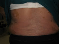
Erythema ab igne


Interesting article from NEJM. It is a common MRCP question for Part 1 & 2a. Do not forget its relationship with hypothyroidism.

This patient has L knee pain.

This reticular, reddish-brown, pruritic, nontender, macular, nonblanching discoloration around the medial aspect of the left knee, with a few superficial erosions, is most consistent with erythema ab igne. This patient had repeatedly applied a heating pad to his left knee in the preceding weeks to relieve discomfort from osteoarthritis.

Read More: New Engl J Med 356;9:e8

Wednesday, July 30, 2008

Optic atrophy

Look for:
  • in young patient, mention you like to examine the eyes for internuclear ophthalmoplegia and cerebellar sign for multiple sclerosis
  • Signs of Cushing's syndrome in SLE pts on steroid
  • in old patient, look for evidence of vascular diseases such as prominent temporal artery (or old scar indicating temporal artery biopsy) and carotid bruit

  • (or endarterectomy scar)


Congenital

If congenital, it is usually hereditary with an onset of deterioration in childhood and may be accompanied by nystagmus. Leber's Hereditary Optic Neuropathy, (LHON) or Leber Optic Atrophy is hereditary, but typically has its onset in 20-30 year old males. This is due to a mutation of the mitochondrial genome and hence is passed exclusively through the mothers. Dominant optic atrophy or Kjer's optic neuropathy has autosomal dominant inheritance. It usually presents in early childhood. There are numerous less common genetically related syndromes.[2]

Alternatively, congenital optic atrophy can be caused by a lack of oxygen during pregnancy, labour or in the early days of a child's life. Some drugs taken during pregnancy are also associated with optic atrophy.


    Acquired


The acquired type of optic atrophy may be due to blood supply changes in the eye or optic nerve (anterior ischemic optic neuropathy or posterior ischemic optic neuropathy), may be secondary to inflammation or swelling within the optic nerve (optic neuritis), may be a result of pressure against the optic nerve (such as from a tumour), or may be related to metabolic diseases (e.g., diabetes mellitus), trauma, glaucoma, or toxicity (caused by methanol, tobacco, or other poisons). It is also seen in vitamin B12 deficiency and Paget's disease of the bone.

Wednesday, July 23, 2008

Proximal myopathy


The clinical diagnosis of proximal myopathy is usually straightforward. Proximal muscle wasting and weakness is easily demonstratable.

Tips:
**The weakness is bilateral and usually symmetrical.
**The sensory is always INTACT!

The causes that you need to consider are:
1. Muscular dystrophy
**Duchenne/ Becker muscular dystrophy (look for pseudohypertrophy of calf muscle
** Fascioscapulohumeral dystrophy (look for facial muscle weakness)
**Limb girdle muscular dystrophy

2. Inflammatory muscle disease

** Polymyositis (muscle may be tender)
** Dermatomyositis (Facial heliotrope rash and Gottron's sign)

3. Endocrinopathy
** Cushing's syndrome
** Thyrotoxicosis
** Acromegaly
**ALL will have obvious external features

4. Metabolic myopathies
** Hypo/hyperkalemia, Hypo/hypercalcemia


The above patient with proximal myopathy also has features of Cushing's syndrome. Remember that Cushing's syndrome can cause proximal myopathy but also do not forget that steroid use for polymyositis/dermatomyositis can also cause Cushing's syndrome.


How do you differentiate polymyositis, dermatomyositis and inclusion body myositis ?

Polymyositis Dermatomyositis Inclusion body myositis


Sex F>M F>M M>F

Age Adult Childhood & Adult Elderly > 50 years old

Rash Yes No No

Distribution of weakness Prox > Distal Prox > Distal Prox = Distal,

predilection for

finger/wrist flexion,

knee extensors

CK incr (up to 50x) N or incr (up to 50x) N or mildly incr (<10

x N)

Muscle biopsy amyloid deposits

Response to

immunosupressive Yes Yes No or minimal

EMG myopathic myopathic myopathic/neuropathic

Monday, July 21, 2008

PACES course 13/14 Sept 2008

This picture was the first picture that came out when I typed PACES in google. Well, I guess it just means that only a few steps and you'll also be there :)

Thanks for the overwhelming response for the PACES course registration. All the seats for the PACES course are filled up.

I am also very happy as I have got several professional tutors who have confirmed coming for the course eg. Prof Richard, Prof Amir, Prof Malik, Prof Raymond, Dr. Haniffah, Prof Rashid, Dato' Dr. Chandran, ....many others who will surprise you on that day.

There are some who have asked regarding the attire on that day. It would be formal and I would advise you to wear as though you will be going for the PACES exam(well, don't worry coz the whole place is air-conditioned)

Thursday, July 17, 2008

Leprosy (Hansen's Disease)


Examine this patient's feet

Look at this patient's face and examine him

You might this case in Malaysia PACES center, station 5.

This patient has leonine facies (face features resemble a lion) and the ear lobe is thickened as well.

Note: The other differential for leonine facies is cutaneous T cell lymphoma.

There is presence of amputated R index finger and L 4th and 5th toe. The small muscles of the hands are wasted.

There is evidence of sensory peripheral neuropathy and the ulnar and common peroneal nerves are thickened on palpation.

The overall picture shows that this patient has lepromatous leprosy complicated by severe neuropathy.

Questions:

1) How would you classify leprosy ?

According to Madrid classification system, it is classified into Tuberculoid, Bordeline and Lepromatous type.

However, WHO classify it into 2 types i.e. paucibacillary and multibacillary type for treatment purposes.

2) How would you investigate this patient ?

- Slit skin smear, skin biopsy

- Nerve conduction study for neuropathy as in this patient

3) How would you manage this patient ?

Start anti Leprosy agents - Clofazamine, Dapsone, Rifampicin

Screen other family members

Tuesday, July 15, 2008

Carotenaemia


I saw this patient in the Medical clinic yesterday.
He is a 45 years old man with Follicular lymphoma who completed chemotherapy CHOP in 2006 with no B symptoms and no enlarged lymph nodes.
If you see him, you would think that he has jaundice. But, his sclera does not show that .

Further history reveals that he has been taking 3 carrots daily since after the chemo as was told to be good from his friends. Yup ! Carotenaemia.

What causes carotenaemia?
Excessive intake of carrots, and/or other yellow and green vegetables and citrus fruits are the usual cause of
carotenaemia (American spelling carotenemia). Carotene is the chief precursor of Vitamin A and is converted to
this in the mucosal cells that line the small intestine. Pancreatic lipase enzymes, bile salts, fat, and thyroid
hormone aid conversion and absorption. Thus carotenaemia is more likely in some diseases such as liver disease,
hypothyroidism and diabetes mellitus where this is impaired. In rare cases, a genetic defect in carotene
metabolism may also be a cause of carotenaemia, without requiring excessive intake of carotene.
Who gets carotenaemia?
Carotenaemia can occur at any age but is most common in young children fed large amounts of commercial
infant food preparations. These foods often contain carrots, pumpkin, squash, spinach and sweet potato, all of
which are high in carotene. Cooking, mashing and pureeing these foods make carotene more available for
absorption. Carotenaemia has also been found in vegetarians or food faddists who over-indulge in carrots and
oranges.
What are the clinical features?
Carotenaemia is characterised by yellow discolouration of the skin, particularly in areas where the horny layer is
thickened such as the soles and palms. It is also most evident on areas where subcutaneous fat is abundant. The
sclera (white outer coating of the eyeball) and mucous membranes (eyes, mouth, nostrils etc) are unaffected. The
presence of yellowing of the sclera usually means there is increased circulating bilirubin and is known as jaundice.

2nd MRCP PACES Preparatory Course


The 2nd MRCP PACES Preparatory Course is finally in place. The organise, wuchereria would like to take this opportunity to welcome participation from all.

Click here to download the course brochure (great design by wuchereria :) & the registration form.

The link will also be mirrored here.

If sending by mail, please address it to:

C/O Professor Richard Loh/ Dr TT Lim
2nd MRCP PACES Preparatory Course
Penang Medical College
No 4 Sepoy Lines,
10450 Penang
Malaysia

Monday, July 14, 2008

Erythema nodosum


Very frequent MRCP exam question, especially Part 1 & 2a, don't be surprised if it comes out in Station 5 PACES.

The picture shows erythematous nodules over the legs involving the shin. They are tender and elevated.
My diagnosis is that this patient has erythema nodosum.

You need to know the causes !

May even appear in history taking if you read Mir &Ryder PACES
Idiopathic/ unknown
Sarcoidosis
Streptococcal infection
Tuberculosis
Infections other than TB or strep - HIV...
Pregnancy or oral contraceptives
Drugs other than OCAs
Inflammatory bowel disease
Behcet's disease
Other

Dermatomyositis


Skin station - common MRCP question. Also seen in Undergraduate medical exams. I remember seeing my first dermatomyositis patient in Sarawak General Hospital when I was a medical student in 3rd year and was told by the physician to remember the characteristic facies.

This patient has heliotrope rash(named after the above flower which I took the photo while I was in UK for my MRCP exam). He also has gottron's papules with proximal myopathy. There are no oral ulcers or arthritis to suggest mixed connective tissue disease. There is a biopsy scar at his R biceps.

What are the investigations you would perform ?
CK, EMG, muscle biopsy, anti Jo1

What is the definitive treatment ?
High dose steroids 1 mg/kg

What is the association ?
Internal malignancy eg NPC, GI, Breast, Ovary, Lung, mostly detected within 2 years

Sunday, July 13, 2008

Marfan syndrome


Always kept for exams for both medical students and MRCP exams.
Always look for this in the CVS station so you'll never miss it. It may also come out in Station 5 for MRCP candidates.

This patient is tall and has presence of arachnodactyly. There is positive wrist sign and steinberg sign. There is also presence of collapsing pulse and corrigan's sign. There is no evidence of disloaction of the lens. He has high arched palate and the chest does not show pectus excavatum or carinatum. There is no stigmata of IE.
The JVP is not raised and there is no pedal oedema. There is no scar on the precordium. The apex is displaced at 6th ICS at L MCL. No parasternal heave, no thrills. Normal 1st and 2nd HS. There is a Grade 3 EDM @ LSE best heard on expiration and sitting forward.

My diagnosis is that this patient has Moderate AR 2 to Marfan syndrome. I would like to look for other signs of AR eg Quincke, de Musset, pistol shot....

What's your differential diagnosis ?
Homocystinuria

What other valve lesions would you suspect in Marfan syndrome patients ?
MVP

What is the inheritance ?
AD

What investigations would you do ?
ECG, CXR, Echo

What is the Echo criteria for valve replacement in AR pt ?
symptomatic, LV end systolic diameter >5.5 cm or LVEF < 50%

If you are allowed to do one test for a patient with AR without Marfan, what test would it be ?
VDRL

Saturday, July 12, 2008

Graves Disease

Looks easy ? If a case looks easy in MRCP, always think again and do something to impress the examiners.

Examination :
Hands - Tremor, onycholysis, Acropashy, Warm sweaty palms
Pulse rate, Irregular to suggest AF ?

Proximal myopathy

Eyes - Exopthalmos, proptosis, Lid lag, lid retraction, Extraocular movements, chemosis

Neck - Scar, Goitre - Inspect, palpate for ? single nodule, multinodular, diffuse(most likely diffuse in Graves), percuss for retrosternal extension, auscultate for bruit

Limb - Pretibial myxedema, tendon reflexes

Well, if you got all those, I guess medical students can do all that.

So, you need to mention whether the disease is active or not.
Signs of activity - tremor, warm&sweaty palms, Thyroid bruit, Tachycardia

So mention the diagnosis as Graves disease and currently the disease is active as evidence by.....

Friday, July 11, 2008

Peutz-Jeghers syndrome


I have never seen this till my practice for the MRCP exam. Well, there are some cases where you will never see before in your whole life except from atlas or websites and it appears for the first time during your MRCP examination. Don't get drawn back. Always stay calm and show the examiner that it is an easy case and talk confidently although you know nuts about it. Remember that the examiner cannot ask you much in the PACES exam because time is short and remember that they are physicians and not dermatologists !

There are small pigmented macules on the lips and buccal mucosa, suggestive of Peutz-Jeghers syndrome. I would like to ask the patient for any abdominal pain or previous history of GI bleed. I would also like to ask for family history of similar problem as it is autosomal dominant.

Peutz-Jeghers is associated with intestinal polyposis which are hamartomas and there is increased incidence of breast, ovarian and pancreatic malignancy.

Other differential diagnosis for this type of hyperpigmentation would include Addison's disease.

Thursday, July 10, 2008

Psoriatic arthropathy


If you think you see RA in MRCP exam, think again ! It may be Psoratic arthropathy RA like or it may also be mixed connective tissue disease.


Examine this patient's hands

This patient has psoriatic arthropathy (Rheumatoid arthritis like) as evidence by bilateral symmetrical deforming arthropathy involving the MCP and PIP joints bilaterally sparing the DIP joints. There is also erythematous plaques with silvery scales involving the hands, scalp, behind the ears, extensor surface of the legs and the trunk. There are also nail changes if pitting and subungal hyperkeratosis but no discolouration,ridging or onycholysis. There is a vertical scar over the R knee suggestive of R knee replacement. There is no evidence of Koebner phenomenon over the scar. There is limitation of movement of the hands but functionally she could still grip and hold a key.


What is this type of psoriasis and what are the other types ?

This is chronic plaque psoriasis and the others include guttate, inverse, erythrodermic, pustular psoriasis


What are the arthropathy of psoriasis ?

- RA like

- Oligoarthritis

- Distal asymmetrical

- Ankylosing spondylitis type

- Arthritis mutilans


How would you like to manage this patient ?

Multidisciplinary approach

Treat pharmacologically and non-pharmacologically

Pharmacologically - Systemic treatment in view of arthropathy and also skin involvement of > 70% eg Methotrexate, Coal tar for the scalp

Non-pharmacologically - physiotherapy, occupational therapy, avoid precipitating factors eg drugs - beta blockers, qunidine, lithium, stress,


What infections can precipitate psoriasis ?

Streptococcal

HIV


Beau's line

I have stressed this to medical student before that nails are very important and in fact you can come to a diagnosis based on nails.
This is one of my patient in the haematology ward who came for chemotherapy for ALL(Acute lymphoblastic leukaemia).

The above shows Beau's line which is due to temporary arrest of nail growth from chemotherapy agent for the haematological malignancy.

Review from http://www.aafp.org/afp/20040315/1417.html

Nail Findings and Associated Systemic Conditions
Nail finding
Associated systemic conditions
Shape or growth change
Clubbing Inflammatory bowel disease, pulmonary malignancy, asbestosis, chronic bronchitis, COPD, cirrhosis, congenital heart disease, endocarditis, atrioventricular malformations, fistulas
Koilonychia Iron deficiency anemia, hemochromatosis, Raynaud's disease, SLE, trauma, nail-patella syndrome
Onycholysis Psoriasis, infection, hyperthyroidism, sarcoidosis, trauma, amyloidosis, connective tissue disorders
Pitting Psoriasis, Reiter's syndrome, incontinentia pigmenti, alopecia areata
Beau's lines Any severe systemic illness that disrupts nail growth, Raynaud's disease, pemphigus, trauma
Yellow nail Lymphedema, pleural effusion, immunodeficiency, bronchiectasis, sinusitis, rheumatoid arthritis, nephrotic syndrome, thyroiditis, tuberculosis, Raynaud's disease
Color change
Terry's (white) nails Hepatic failure, cirrhosis, diabetes mellitus, CHF, hyperthyroidism, malnutrition
Azure lunula Hepatolenticular degeneration (Wilson's disease), silver poisoning, quinacrine therapy
Half-and-half nails Specific for renal failure
Muehrcke's lines Specific for hypoalbuminemia
Mees' lines Arsenic poisoning, Hodgkin's disease, CHF, leprosy, malaria, chemotherapy, carbon monoxide poisoning, other systemic insults
Dark longitudinal streaks Melanoma, benign nevus, chemical staining, normal variant in darkly pigmented people
Longitudinal striations Alopecia areata, vitiligo, atopic dermatitis, psoriasis
Splinter hemorrhage Subacute bacterial endocarditis, SLE, rheumatoid arthritis, antiphospholipid syndrome, peptic ulcer disease, malignancies, oral contraceptive use, pregnancy, psoriasis, trauma
Telangiectasia Rheumatoid arthritis, SLE, dermatomyositis, scleroderma

Tuesday, July 8, 2008

Tendon xanthomas

This is a common MRCP exam question. My friend just got this 1 week ago when sitting for the exam in Singapore.

There are 2 differential diagnosis that you have to think about
1) Chronic tophaceous gout
2) Tendon xanthomas

The question is how to differentiate both of them. One simple test is that tendon xanthomas move with the tendon. So ask the patient to move the finger and feel the lesions and see whether it moves with the tendon.
Look also at the distribution of the deposits, in tendon xanthomas over the knuckles, elbows, knees, Achilles tendon. Look also for xanthalesma.
For gouty tophi, look at the ear lobes, feet, elbows.

Comment
Tendon xanthomas are seen in 75% of adults with familial
hypercholesterolaemia. They are intra-and extra-cellular
accumulations of cholesterol, noticed on extensor
tendons over knuckles, achilles, knee, and elbows. They
decrease with treatment of hypercholesterolaemia. They
are also seen in familial defective apo B100, type 3
hyperlipoproteinaemia, and sitosterolaemia

Reference
1. Mahley RW, Weisberger KH, Farese RV. Disorders of Lipid
Metabolism in Wilson JD, Foster DW, Kronenberg HM, Larsen
PR (eds). William’s Textbook of Endocrinology (9th ed).
Philadelphia, WB Saunders 1998; 23: 1099-1153.

Monday, July 7, 2008

Systemic lupus erythematosus

For MRCP PACES candidates,
besides Psoriasis, please remember this as one of the most common exam questions.
I got this case in my 3rd attempt of PACES in Malaysia.

This patient has a malar rash. Describes as erythematous rash over the cheeks and bridge of nose and sparing the nasolabial folds. There is also presence of hypo and hyperpigmentation, however there is no follicular plugging.
My diagnosis is that this patient most likely has SLE and I would proceed to look for oral ulcers, discoid lupus, arthritis, alopecia, listen to the heart for pericardial rub, listen to the lungs for pleural effusion(serositis) and examine the abdomen for presence of hepatosplenomegaly. I would also like to look at the fundus for cytoid bodies.

Criteria for SLE
1) Malar rash
2) Discoid lupus
3) Photosensitivity
4) Oral ulcers
5) Arthritis
6) Serositis (Pleuritis/Pericarditis)
7) Renal disorder : Proteinuria >500 mg/d or cellular casts
8) Neurological disorder
9) Haematological disorder
10) Immunological disorder : anti dsDNA, anti Smith Ab, ACA/LA, false positive VDRL
11) ANA

Need 4 out of 11 criteria

What investigations would you perform to look for activity ?
ESR, C3, C4, CRP

What is the most common neurological manifestation ?
Psychosis

Saturday, July 5, 2008

Splinter haemorrhage


For medical students, always start examination from the periphery unless told not too in a short case. The nail is one of the most important clue. We saw clubbing previously and this is the diagram of a splinter haemorrhage.
The most common cause for splinter haemorrhage is trauma. Yes, it is not infective endocarditis. But you must not forget IE as one of the cause.

Can any medical students tell me in 'comments' other signs of infective endocarditis ?

The answer is
Osler nodes, Janeway lesion, Roth spot, clubbing.

Worth remembering !

Modified Duke criteria

Pathological criteria

  • Positive histology or microbiology of pathological material obtained at autopsy or cardiac surgery (valve tissue, vegetations, embolic fragments, or intracardiac abscess content)

Major criteria

  • Two positive blood cultures showing typical organisms consistent with infective endocarditis, such as Streptococcus viridans and the HACEK group OR
  • Persistent bacteraemia from two blood cultures taken > 12 hours apart or three or more positive blood cultures where the pathogen is less specific, such as Staphylococcus aureus and Staph epidermidis OR
  • Positive serology for Coxiella burnetti, Bartonella species, or Chlamydia psittaci OR
  • Positive molecular assays for specific gene targets
  • Positive echocardiogram showing oscillating structures, abscess formation, new valvular regurgitation, or dehiscence of prosthetic valves

Minor criteria

  • Predisposing heart disease
  • Fever > 38°C
  • Immunological phenomena such as glomerulonephritis, Osler's nodes, Roth spots, or positive rheumatoid factor
  • Microbiological evidence not fitting major criteria
  • Elevated C reactive protein or erythrocyte sedimentation rate
  • Vascular phenomena such as major emboli, splenomegaly, clubbing, splinter haemorrhages, petechiae, or purpura

Definite infective endocarditis

  • Pathological criteria positive OR
  • Two major criteria OR
  • One major and two minor criteria OR
  • Five minor criteria

Wednesday, July 2, 2008

Fundoscopy - DM proliferative retinopathy

This is also one of the commonest MRCP exam question. Even medical students should know how to look at this.

When you reach this station, look around.
If you see a walking stick - Retinitis pigmentosa
If you see diabetic dermopathy- DM retinopathy(usually proliferative)
If you see an infusion pump - Grade 4 hypertensive retinopathy
If you see malar rash or Cushingoid facies - Optic atrophy

Sounds easy......but still one of the most feared exam station in PACES

The patient has DM proliferative retinopathy as evidence by multiple panphotocoagulation scars at the peripheral and also there is neovascularization. There are also dot blot haemorrhages and soft exudates.