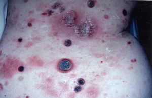
 I am sure that this is possibly seen during the MRCP PACES exam. I have managed to see a few of these patients while travelling through the journey in MRCP and I could remember my friend who showed me this while patient was in the skin ward. Unfortunately the patient died a few days later.
So, could anyone tell me what is the diagnosis ?
Cutaneous T-cell lymphoma(CTCL is a form of T cell lymphoma first manifested in the skin, but because the process involves the entire lymphoreticular system, the lymph nodes and internal organs become involved.
Another term - mycosis fungoides (nothing to do with fungus)
Plaques, scaling or non-scaling, large(>3 cm) at superficial
(looks like eczema or psoriasis initially), later it becomes thicker and "infiltrated"
Nodules and tumours with or without ulcer
Poikiloderma with or without plaques and nodules
Extensive infiltration can cause leonine facies
Often spares exposed areas initially
Checklist of diagnosis :
1) History
2) Lymph node - biopsy if palpable
3) Skin biopsy with 1 micrometer sections
4) CXR
5) PBF
6) CT scan abdomen
7) Buffy coat - abnormal circulating T cells (Sezary type) and increased WBC (20,000)
Management :
pre CTCLor proven plaque stage with no lymphadenopathy/criculating T cells - PUVA photochemotherapy in most effective, topical chemotherapy with notrogen mustard in ointment base(10mg%)
If isolated tumours develop, should be treated with local x-ray or electron beam therapy
Extensive plaque stage - electron beam plus chemotherapy
Source :
- www.onexamination.com
- Color atlas and synopsis of Clinical Dermatology by Thomas B. Fitzpatrick et. al. (McGraw Hill)

11 comments:
From the inspection, 1 can notice erythematous macules, hemorrhagic lesions with central necrosis, 2 crusted lesions suspected of rupture of bullae on his lower back and buttocks are suggestive of meningococcemia.
Differential Diagnosis:
Erythema Multiforme.
Steven Johnsons Syndrome
Sorry, not correct. Any other guesses?
Ok, I was asked to give a clue via sms. I have also seen a case while doing Haematology posting.
Warfarin induced skin necrosis
IMHO,isn't cutaneous antrax?
CUTANEOUS T- CELL LYMPHOMA.
Great ! Yea. The answer is CTCL.
Hi just wondering,what are the points in that picture which tell that this is a CTCL,is there a specific sign?Cause all i see is multiple hemorrhage and some bullae...
i see..so long time not log in to read this blog already. Finally, i can graduate as a new HO already..
can any dr tell me why is it Cutaneous T-cell Lymphome
CTCL is not easy to diagnose. Initially patients may present with lesions looking like psoriasis or eczema.
The stages include
Pre-mycosis fungoides
Patch stage
Plaque stage
Tumour stage
The picture shows area of erythematous patch and also areas of plaque. THere is also existing tumour becoming huge or mushroom shaped. Lesions has become necrosis and ulcerated.
Anyway, if you see a person who looks erythrodermic with lesions that look like being burned, always think of CTCL.
Check for lymphadenopathy and also PBF to look for Sezary cells.
They don't respond to treatment used to treat psoriasis or eczema....except maybe PUVA.
Subsequently the biopsy is the way to diagnose.
Post a Comment