Tuesday, March 31, 2009

Pulmonary embolism


Case by Chye Chung 

Thanks for the case which is a good case to discuss.

28 years old Malay man who is in an army, presented with sudden onset of shortness of breath and localised pleuritic chest pain for 1 day. He also has non-productive cough and with an episode of haemoptysis, low grade fever without chills and rigors and rashes. He had previous history of trauma to the knww and ACL repair done 2 months ago. No history of pneumonia, PTB, recent air travel or long journey drive. No other medical illness. 

Physical examination was unremarkable and lungs clear with no loud P2

D-dimer 300
Plt 409
PT12.9, aPTT 29.2 INR 1.07
pH7.469, pCO2 72.0, HCO3- 25.8, BE 3.3 O2 sat 95.4%
CXR normal

Would anyone like to comment on the ECG and CTPA ?

Thanks again to Chye Chung for the post !

Sunday, March 29, 2009

ECG Quiz

What abnormality is shown ?

What is the underlying problem ?

What is your management ?

Saturday, March 28, 2009

Quiz


What sign is shown here ?

Where is the lesion ?

What are the possible causes ?

What investigation can you perform ?


Friday, March 27, 2009

Quiz Skin


What is the diagnosis and why ?

What are the complications ?

What is the treatment ?


Thursday, March 26, 2009

Behcet's Disease


Sourece : www.dermis.net

I saw a patient in the ward today whom my MO told me that she had ulcers in the mouth and genitalia. One of the diagnosis that came to my mind at that time was this. Common in MRCP PACES part 1 and 2a. Not common in PACES MRCP.

What is the diagnosis ?

What test can be done to support the diagnosis ?

What is the diagnostic criteria ?

What is the treatment ?

Hulusi Behcet was born on Feb 20 1889 in Istanbul. As Professor and Director of Department of Dermatology in Istanbul University, he saw the first case of his disease with oral apthous ulcers, genital ulceration, erythema nodosum and visual loss in 1924. The patient was diagnosed as tuberculosis or syphilis, but his diagnosis was viral infection. He died of heart attack on March 8 1948.

Behcet disease is a recurrent systemic inflammatory disorder of unknown aetiology.
It was first described in 1937 as a triad of recurrent oral ulceration, genital ulceration and uveitis. 

Males are more common, usually in the 3rd decade.
Associated with HLA B-51
Common in Turkey, Mediterranean and Japan

Diagnostic criteria - International Study Group for Behcet's Disease 1990
Recurrent oral aphthae - at least 3x in a year
PLUS 2 of
  • Genital ulceration - active lesion/scar
  • Skin lesion - erythema nodosum, foliculitis, other ulcerations
  • Eye involvement - anterior or posterior uveitis, or retinal vasculitis
  • positive pathergy test - skin hyperreativity to pin prick( sterile pustule formed in 24 - 48 hours)
Treatment involves Steroids, cyclosporin, cyclophosphamide or azathioprine.
Colchicine may be effective in treating ulceration.

Wednesday, March 25, 2009

Superior Vena Cava Obstruction


I still remembered during my 2nd year as medical student that one of my lecturer who was an Anaesthesiologist(Dr. Murthy) who took us for Respiratory system showed us one case in Sarawak General Hospital.
His words : Look at this patient and remember this forever because you may not see it for a long time to come

He performed the Pemberton sign by asking the patient to lift his arms and immediately, the patient's face became plethoric and his voice became rather hoarse that he had to immediately put down his arms.

You may be asked to examine the respiratory system and you'll find
1) Dilated veins over the chest
2) Plethoric face, occasionally with congested eyes
3) Non-pulsatile raised JVP
4)Pemberton sign positive (you may not want to perform this but offer to the examiner because you may distress the patient which will then cause you to fail)
5) Talk to the patient to notice the hoarseness of voice

You are also expected to find the cause for this which almost always caused by lymph node compression due to malignancy. 
The above patient has Non-Hodgkin's lymphoma with SVC obstruction and is in respiratory distress.


Monday, March 23, 2009

Argyll Robertson pupil


This is an interesting video I downloaded from youtube. I am sure it will be benefeficial for medical students to understand this.

I used to have problem remember when the pupil constricts and when it dilates till I heard this joke :
A prostitute will accomadate but will not react.

So in Argyll Robertson pupil(due to Neurosyphilis), the pupil will constrict to accomadation reflex but will not constrict to light - just as what the video showed.
It is due to damage to the periaqueductal area and is a classic sign.
The pupil is small, irregular and fixed to light but reactive to accomodation
Other causes of what perhaps incorrectly called AG pupils are pinealomas, diabetes, brainstem encephalitis, multiple sclerosis and Wernicke's encephalopathy. These conditions cause fixed semidilated pupils and sometimes unequal pupils; only a small, irregular, fixed pupil should be attributed to neurosyphilis.

I was fortunate, well unfortunate for the patient to have managed a case of neurosyphilis 2 years ago in Neurology . I still remembered the LP I did for the patient (as above) which showed VDRL positive in the LP. Patient actually improved after treated with Penicillin.

Now, the question I want to ask is - what if you get pupils which react to light but not to accomodation (reversed Argyll Robertson pupil). What is the cause ?
Clue : It occured in 1920s


Quiz


I am sure you know the answer if you are from Malaysia.

What is the diagnosis and what is the classical description ?


Friday, March 13, 2009

Cutaneous T cell lymphoma

I am sure that this is possibly seen during the MRCP PACES exam. I have managed to see a few of these patients while travelling through the journey in MRCP and I could remember my friend who showed me this while patient was in the skin ward. Unfortunately the patient died a few days later.

So, could anyone tell me what is the diagnosis ?

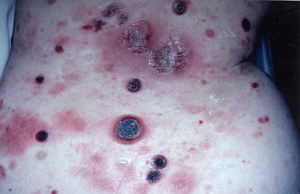
Cutaneous T-cell lymphoma(CTCL is  a form of T cell lymphoma first manifested in the skin, but because the process involves the entire lymphoreticular system, the lymph nodes and internal organs become involved.
Another term - mycosis fungoides (nothing to do with fungus)

Plaques, scaling or non-scaling, large(>3 cm) at superficial
(looks like eczema or psoriasis initially), later it becomes thicker and "infiltrated"
Nodules and tumours with or without ulcer
Poikiloderma with or without plaques and nodules
Extensive infiltration can cause leonine facies

Often spares exposed areas initially

Checklist of diagnosis :

1) History
2) Lymph node - biopsy if palpable
3) Skin biopsy with 1 micrometer sections
4) CXR
5) PBF
6) CT scan abdomen
7) Buffy coat - abnormal circulating T cells (Sezary type) and increased WBC (20,000)

Management :
pre CTCLor proven plaque stage with no lymphadenopathy/criculating T cells - PUVA photochemotherapy in most effective, topical chemotherapy with notrogen mustard in ointment base(10mg%)

If isolated tumours develop, should be treated with local x-ray or electron beam therapy
Extensive plaque stage - electron beam plus chemotherapy

Source :
  • www.onexamination.com
  • Color atlas and synopsis of Clinical Dermatology by Thomas B. Fitzpatrick et. al. (McGraw Hill)


Thursday, March 12, 2009

Chronic liver disease

The above picture shows gynaecomastia and multiple spider naevi. 
When you see this in the exam whether medical student or MRCP, think of none other than chronic liver disease (liver cirrhosis)
You need to know the signs at your finger tips.
Nails - leuconychia, clubbing
Wrist/Hands - flapping tremor, palmar erythema, Dupuytren's contracture
Face - Jaundice, Parotid swelling(esp alcoholic), look also for KF ring(Wilson)
Chest - Gynaecomastia, spider naevi, loss of axillary hair
Abdomen - Caput medussa, prominent veins, Ascites, Splenomegaly
Legs - Oedema

Note that you rarely get hepatomegaly in chronic liver disease because by then the liver would be cirrhotic and shrink. However, if it is enlarged, think of malignant transformation or alcoholic liver disease.

Can you list the investigations you would perform in these patients ?