Tuesday, October 6, 2009

Clubbing


It's been some while since I last wrote. I have been busy with Neurology. Well, this patient is a 60 years old Malay man who was admitted to my ward with progressive L sided weakness over a week. He is non toxic looking and there is no stigmata of chronic liver disease. He has been a chronic smoker. To tell the truth, I was rather upset that 2 of my house officers could not come to a conclusion even after I mentioned the gross clubbing in this patient.
I hope medical students reading this will practice what has been learned in medical student years.
This is a clear cut case of Ca lung with metastasis to the brain.
What I would like to stress here is - Do not forget the causes of clubbing !

Respiratory :
Ca lung
Pulmonary fibrosis
Bronchiectasis
Chronic suppurative lung diseases
Cystic fibrosis

CVS :
Congenital cyanotic heart disease
Eisenmenger syndrome
Infective endocarditis

GI :
Chronic liver disease (Cirrhosis)
Inflammatory bowel disease

Thyrotoxicosis (pseudoclubbing - acropachy)

Monday, June 22, 2009

Plantar Response - Other than Babinski

art-sin447785.fig1.gif

Other than Babinski sign, what other signs do you know ?

In upper motor neuron lesion, the big toe will dorsiflex with the following :

Gordon reflex – On applying pressure to the muscle of the calf

Oppenheim sign – on applying heavy pressure with thumb and index finger to the shin, stroking downwords from below the knee down to the ankle

Bing reflex – when the dorsum of the toe is pricked with a pin

Schaefer reflex – pinching the Achilles tendon sufficiently to cause pain

Chadock reflex – on stroking the lateral side of the foot, beginning below the malleolus and extending anteriorly along the dorsum of the foot to the base of the big toe

Gonda reflex – grasping the small toes between the fingers, slowly and forcibly flexing the toe and then suddenly releasing the toe


I know an MRCP examiner who likes to ask this question. Try practicing it !  Physical examination is the most crucial step in Neurology.


Saturday, June 6, 2009

Infectious Disease for MRCP Part 2a


39 years old 39 years old Malay Man presented with fever for 1 week associated with cough, yellowish sputum, pleuritic chest pain. No haemoptysis, loss of weight or night sweats. Chronic smoker. Also has diarrhoea for 1 week. 

Examination reveals clubbing, bibasal crepitations, hepatosplenomegaly. Multiple L/N enlargement.

Hb 10

WBC 4.1

Plt 43

Na 112 K3.2 U 3.8 Cr 69


pH 7.43 pCO2 2.4 pO2 9.8 HCO3 12 BE -10 SpO2 95%


What is your choice of treatment ?

A. IV Ceftazidime 2 g stat and 8H

B. IV (sulfamethoxazole and trimethoprim)Bactrim 3 ampules stat & qid

C. Isoniazid, Rifampicin, Ethambutol and Pyrazinamide

D. IV Acyclovir 500 mg stat and 8H

E. IV Amphotericin B 

Tuesday, May 26, 2009

MRCP PACES Course 2009

The time we feared most and the time we can't wait for it to be over. However, we choose to face it. PACES exam.....
We know that part of it is due to pure luck but if you are better prepared, the chances of passing is much higher.
Once again, Penang General Hospital, Seberang Jaya Hospital and Penang Medical College is planning for a MRCP PACES course the August 29 and 30. This is a soft launch for the course for those whom I am grateful for keeping up this blog. 

Thank you very much for the support. All entries are now closed. 

All the best !!


Friday, May 22, 2009

Neurology Quiz


(Archieves of Neurology Vol 66 (No 4) Apr 2009

What is the diagnosis and what is the differentials for this ?


Thursday, May 21, 2009

Tuberculous meningitis


A 45 year old man was admitted with a 3 day history of coma following 2 weeks of headache and fever. Examination revealed a GCS of 8/15, neck stiffness, and a complete R oculomotor nerve palsy. Examination of the CSF showed an opening pressure of 20 cmH2O, 800 leucocytes.mm3 (30% neutrophils, 70% lymphocytes), protein 180 mg/dl, CSF/ blood glucose 0.18. Gram and Ziehl-Neelsen stains of the CSF were negative. Culture of the CSF on Lowenstein Jensen media grew Mycobacterium tuberculosis.
(Adapted from Practical Neurology 2002)

This type of scenario can come out in the Part 1 or 2a MRCP and medical student exams. Not possible for PACES.

Name 3 complications of TB meningitis that may need neurosurgical intervention.


Sunday, May 10, 2009

Meningitis


Photo taken form www.thereddragonhood.com/ images/rabbit.jpg

Text adapted from Archieves of Neurology (April 2009)

A 21 years old previously healthy man presented with 7 days cough with black and yellowish sputum, fever, chills, myalgia and pleuritic chest pain. 3 days before admission, had progressive worsening headache with neck stiffness and 8 episodes of watery diarrhoea

He had taken amoxycillin for 1 day

He had recently changed job, working as a professional landscaper 7 days before onset of symptoms

He had performed lawn mowing and leaf blowing services and noted he had dead rabbits in the areas he worked

T - 39.6 C, Alert, orientated to time, place, person

Stiff neck, no weakness or numbness

Lungs - bibasal crepts with exp ronchi

No lymphadenopathy,  skin lesions, ulcerations

CT brain - Normal

WBC - 10.4 (78% N) ESR - 40 mm/h

CSF - Cell count 1416/mm3, 19% N 73% L RBC 23, glu 41 mg/dl(glu 123 mg/dl), pro 2660 g/dl

Gm stain CSF -ve

CXR - N

MRI - cerebellar tonsillar herniation consistent with Chiary Type 1 malformation without evidence of brainstem syrinx

Treated with IV Ceftrixone and IV Vancomycin

Day 4 , headache worse and developed diplopia and nausea

Repeated LP -Opening pressure - 49 cmH2O, total nucleated cell count 2590/mm3 32% N 43%L 25% M, glu 28 mg/dL, pro 2950 g/dL (RBS - 128 mg/dL) 

Gram stain negative

What is the diagnosis ?

Monday, April 27, 2009

Parkinson Disease

Picture taken from http://www.goldbamboo.com/pictures-t1604.html

For those taking MRCP PArt 1 Ireland (True of False)

The following increases the risk of Parkinson disease
A. Tea
B. Smoking
C. Coffee
D. Low socioeconomic status
E. Physical inactivity


Excellent Neurology book


Congratulations to Dr. Eow and Dr. Samuel for coming out with this excellent book with colours and lots of algorithm with facts for the MRCP candidates, medical students and trainees of Neurology. A 393 pages book.
If there is anyone who is interested, feel free to contact me at thienthienlim@gmail.com.


Saturday, April 25, 2009

Spinocerebellar Ataxia


Just a stimulus to all inspired doctors, physicians and neurologists. A very touching video from youtube on spinocerebellar ataxia.
A reminder to those sitting for the next PACES exam. If you see a young man or lady with cerebellar signs and pyramidal signs, always think of 
1) Spinocerebellar ataxia
2) Multiple sclerosis - transverse myelitis with cerebellar involvement (classical MS)
3) Friedrich's ataxia


Friday, April 24, 2009

ECG Quiz


This is a 34 years old man who was admitted with a syncopal attack. ECG shows the above. What is the diagnosis and what would you do next.

This question recently came out in the MRCP PACES exam Part IIa.


Saturday, April 11, 2009

Rheumatology/Endocrine quiz


Would anyone want to comment on these photos which I took of a boy when I was working in Casualty?


Friday, April 10, 2009

Vitiligo


This patient has areas of depigmentation around the mouth, the upper limb, legs and trunk. He has vilitigo.

Vitiligo affects 1% of the population. Equal between males and females, although more females present to the skin clinic. Family history in 1/3 of patients.
They have Koebner's phenomenon - lesion appearing on the site of lesion.
When you see a patient like this, look for other features of autoimmune

Thyroid disorder - Hashimoto's thyroiditis, Graves' disease
Pallor - Pernicious anaemia
Pigmentation of buccal, skin crease, hypotension - Addison's disease
Tetany, cramps, paraesthesia, Chvostek's and Trousseau's sign - Idiopathic hypoparathyroidism
Fundus - diabetes mellitus
Lungs (fine crepts) - Fibrosing alveolitis
Jaundice - Chronic active hepatitis
Excoriation marks, xanthalesma, pigmentation - primary biliary cirrhosis
rash, arthritis, prox weakness - SLE, RA, dermatomyositis, polymyositis


Look out for polyglandular autoimmune disease
What are the 2 types ?


Tuesday, April 7, 2009

Skin Quiz


This gentleman complaints of this rash over the face.

Describe the rash.

What is the most likely cause.

What investigation would you do ?

What one question would you ask this patient ?

What treatment would you give to this patient ?

Tuesday, March 31, 2009

Pulmonary embolism


Case by Chye Chung 

Thanks for the case which is a good case to discuss.

28 years old Malay man who is in an army, presented with sudden onset of shortness of breath and localised pleuritic chest pain for 1 day. He also has non-productive cough and with an episode of haemoptysis, low grade fever without chills and rigors and rashes. He had previous history of trauma to the knww and ACL repair done 2 months ago. No history of pneumonia, PTB, recent air travel or long journey drive. No other medical illness. 

Physical examination was unremarkable and lungs clear with no loud P2

D-dimer 300
Plt 409
PT12.9, aPTT 29.2 INR 1.07
pH7.469, pCO2 72.0, HCO3- 25.8, BE 3.3 O2 sat 95.4%
CXR normal

Would anyone like to comment on the ECG and CTPA ?

Thanks again to Chye Chung for the post !

Sunday, March 29, 2009

ECG Quiz

What abnormality is shown ?

What is the underlying problem ?

What is your management ?

Saturday, March 28, 2009

Quiz


What sign is shown here ?

Where is the lesion ?

What are the possible causes ?

What investigation can you perform ?


Friday, March 27, 2009

Quiz Skin


What is the diagnosis and why ?

What are the complications ?

What is the treatment ?


Thursday, March 26, 2009

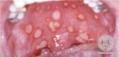
Behcet's Disease


Sourece : www.dermis.net

I saw a patient in the ward today whom my MO told me that she had ulcers in the mouth and genitalia. One of the diagnosis that came to my mind at that time was this. Common in MRCP PACES part 1 and 2a. Not common in PACES MRCP.

What is the diagnosis ?

What test can be done to support the diagnosis ?

What is the diagnostic criteria ?

What is the treatment ?

Hulusi Behcet was born on Feb 20 1889 in Istanbul. As Professor and Director of Department of Dermatology in Istanbul University, he saw the first case of his disease with oral apthous ulcers, genital ulceration, erythema nodosum and visual loss in 1924. The patient was diagnosed as tuberculosis or syphilis, but his diagnosis was viral infection. He died of heart attack on March 8 1948.

Behcet disease is a recurrent systemic inflammatory disorder of unknown aetiology.
It was first described in 1937 as a triad of recurrent oral ulceration, genital ulceration and uveitis. 

Males are more common, usually in the 3rd decade.
Associated with HLA B-51
Common in Turkey, Mediterranean and Japan

Diagnostic criteria - International Study Group for Behcet's Disease 1990
Recurrent oral aphthae - at least 3x in a year
PLUS 2 of
  • Genital ulceration - active lesion/scar
  • Skin lesion - erythema nodosum, foliculitis, other ulcerations
  • Eye involvement - anterior or posterior uveitis, or retinal vasculitis
  • positive pathergy test - skin hyperreativity to pin prick( sterile pustule formed in 24 - 48 hours)
Treatment involves Steroids, cyclosporin, cyclophosphamide or azathioprine.
Colchicine may be effective in treating ulceration.

Wednesday, March 25, 2009

Superior Vena Cava Obstruction


I still remembered during my 2nd year as medical student that one of my lecturer who was an Anaesthesiologist(Dr. Murthy) who took us for Respiratory system showed us one case in Sarawak General Hospital.
His words : Look at this patient and remember this forever because you may not see it for a long time to come

He performed the Pemberton sign by asking the patient to lift his arms and immediately, the patient's face became plethoric and his voice became rather hoarse that he had to immediately put down his arms.

You may be asked to examine the respiratory system and you'll find
1) Dilated veins over the chest
2) Plethoric face, occasionally with congested eyes
3) Non-pulsatile raised JVP
4)Pemberton sign positive (you may not want to perform this but offer to the examiner because you may distress the patient which will then cause you to fail)
5) Talk to the patient to notice the hoarseness of voice

You are also expected to find the cause for this which almost always caused by lymph node compression due to malignancy. 
The above patient has Non-Hodgkin's lymphoma with SVC obstruction and is in respiratory distress.


Monday, March 23, 2009

Argyll Robertson pupil


This is an interesting video I downloaded from youtube. I am sure it will be benefeficial for medical students to understand this.

I used to have problem remember when the pupil constricts and when it dilates till I heard this joke :
A prostitute will accomadate but will not react.

So in Argyll Robertson pupil(due to Neurosyphilis), the pupil will constrict to accomadation reflex but will not constrict to light - just as what the video showed.
It is due to damage to the periaqueductal area and is a classic sign.
The pupil is small, irregular and fixed to light but reactive to accomodation
Other causes of what perhaps incorrectly called AG pupils are pinealomas, diabetes, brainstem encephalitis, multiple sclerosis and Wernicke's encephalopathy. These conditions cause fixed semidilated pupils and sometimes unequal pupils; only a small, irregular, fixed pupil should be attributed to neurosyphilis.

I was fortunate, well unfortunate for the patient to have managed a case of neurosyphilis 2 years ago in Neurology . I still remembered the LP I did for the patient (as above) which showed VDRL positive in the LP. Patient actually improved after treated with Penicillin.

Now, the question I want to ask is - what if you get pupils which react to light but not to accomodation (reversed Argyll Robertson pupil). What is the cause ?
Clue : It occured in 1920s


Quiz


I am sure you know the answer if you are from Malaysia.

What is the diagnosis and what is the classical description ?


Friday, March 13, 2009

Cutaneous T cell lymphoma

I am sure that this is possibly seen during the MRCP PACES exam. I have managed to see a few of these patients while travelling through the journey in MRCP and I could remember my friend who showed me this while patient was in the skin ward. Unfortunately the patient died a few days later.

So, could anyone tell me what is the diagnosis ?

Cutaneous T-cell lymphoma(CTCL is  a form of T cell lymphoma first manifested in the skin, but because the process involves the entire lymphoreticular system, the lymph nodes and internal organs become involved.
Another term - mycosis fungoides (nothing to do with fungus)

Plaques, scaling or non-scaling, large(>3 cm) at superficial
(looks like eczema or psoriasis initially), later it becomes thicker and "infiltrated"
Nodules and tumours with or without ulcer
Poikiloderma with or without plaques and nodules
Extensive infiltration can cause leonine facies

Often spares exposed areas initially

Checklist of diagnosis :

1) History
2) Lymph node - biopsy if palpable
3) Skin biopsy with 1 micrometer sections
4) CXR
5) PBF
6) CT scan abdomen
7) Buffy coat - abnormal circulating T cells (Sezary type) and increased WBC (20,000)

Management :
pre CTCLor proven plaque stage with no lymphadenopathy/criculating T cells - PUVA photochemotherapy in most effective, topical chemotherapy with notrogen mustard in ointment base(10mg%)

If isolated tumours develop, should be treated with local x-ray or electron beam therapy
Extensive plaque stage - electron beam plus chemotherapy

Source :
  • www.onexamination.com
  • Color atlas and synopsis of Clinical Dermatology by Thomas B. Fitzpatrick et. al. (McGraw Hill)


Thursday, March 12, 2009

Chronic liver disease

The above picture shows gynaecomastia and multiple spider naevi. 
When you see this in the exam whether medical student or MRCP, think of none other than chronic liver disease (liver cirrhosis)
You need to know the signs at your finger tips.
Nails - leuconychia, clubbing
Wrist/Hands - flapping tremor, palmar erythema, Dupuytren's contracture
Face - Jaundice, Parotid swelling(esp alcoholic), look also for KF ring(Wilson)
Chest - Gynaecomastia, spider naevi, loss of axillary hair
Abdomen - Caput medussa, prominent veins, Ascites, Splenomegaly
Legs - Oedema

Note that you rarely get hepatomegaly in chronic liver disease because by then the liver would be cirrhotic and shrink. However, if it is enlarged, think of malignant transformation or alcoholic liver disease.

Can you list the investigations you would perform in these patients ?


Thursday, February 26, 2009

Neurology Quiz

1) Present your findings. 


2) What is the diagnosis ?


3) Give reason for your answer.


4) What other differential diagnosis ?


5) What other signs would you look for ?

Thursday, January 29, 2009

Ptosis/Exopthalmos


Can you decide during the PACES MRCP exam whether the R eye is having exopthalmos or the L eye is having ptosis ?
Do you need to toss the coin to decide this in the exam.....50% right, 50% wrong.

If you see the neck you may see a goitre but again...don't be fooled as Graves disease may coexist with myasthenia gravis as both of them are autoimmune disease

One technique to know fast is think logically -
If you see this patient in Station 3(Neuro) - the possibility of L ptosis is higher
If you see this patinet in Station 5(Endocrine) - the possibility of R exopthalmos is higher

So, in Station 3, you would check the eyes as if it was approach to ptosis and if in Station 5 you would check the eyes and look for signs of thyrotoxicosis.

This patient actually has Graves disease.

Tuesday, January 27, 2009

Giant cell arteritis


You will hear this term over and over again from medical school to MRCP PArt 1 to MRCP PArt 2a to PACES(most of the time in history station for differential of headache or transient blurring of vision).

Mean age of diagnosis 70 years old
Slightly more common in women

Symptoms :
Headache - usually localised
Jaw claudication (pain on chewing)
General malaise
Proximal muscle stiffness and pain
Visual loss (Amaurosis fugax)

Signs :
Tender and thickened scalp veins
Optic disc swollen and later pale (AION)

Investigations :
ESR elevated (one of the differential for ESR >100)
Raised CRP
Raised ALP
Superficial temporal artery biopsy - intimal thickening and medial damage, giant cells with inflammatory cell infiltration in the internal elastic lamina (GOLD STANDARD)
Management :
A general treatment regime for GCA patients that many physicians follow begins with a 70mg/day prednisone dose.

After the first week, the dose is reduced by 10mg every week until the dosage reaches 20mg/day (Schmidt, 2006).

However, some physicians recommend that the high initial dose be maintained for two weeks (Rahman & Rahman, 2005).

Once the 20mg/day level is reached, the dosage is lowered by 2.5mg per week.

Then, when the dosage reaches 10mg/day, it is reduced by 1mg every month depending on the symptoms and test results (Schmidt, 2006).

Most patients remain on corticosteroids for 1-2 years, although some may require long-term steroid treatment (Schmidt, 2006; Piptone et al., 2005).

The reason that treatment of GCA is usually short in duration is because GCA is a self-limiting disease for most patients (although the mechanism for this limitation is unknown) (Azhar et al., 2005; Schmidt, 2006).

Longer corticosteroid treatment is typically required for patients with more severe GCA; these patients usually have cytokine levels (such as TNF-α) above most GCA patients (Hernandez-Rodriguez et al., 2004).

[www.bio.davidson.edu]

Monday, January 5, 2009

ECG Quiz

This is a 63 year old Chinese lady who presented with dizziness.


1. What does the ECG show ?


2. What is the treatment ?